
Breast cancer is one of the most common cancers affecting women worldwide, and early detection is one of the most effective ways to improve survival rates. Throughout the years, medical imaging technology has advanced, making diagnosis more accurate and less invasive. Among these advances, 3D mammography and breast MRI developed as powerful tools for early detection. This article explores how imaging methods work, limitations, and combined role developing breast cancer diagnosis. These techniques help in identifying cancer at its earliest stages but also reduce unnecessary biopsies.
How We Detect Breast Cancer
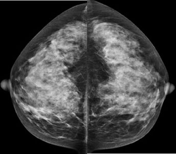
Breast cancer grows when cells are abnormal in the breast and grow wildly and form a tumor. Early detection of these tumors before spreading to other parts of the body is necessary for successful treatment. 2D mammography has been the gold standard for spans, but it has limitations, particularly for those women with dense breast tissue. Dense tissue can hide tumors, making the tumor harder to detect and increasing the risk of diagnoses. That is why, 3D mammography and breast MRI come and offer more clarity and accuracy.
Why is 3D Mammography known as DBT?
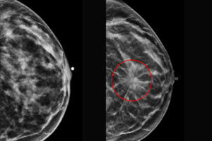
3D mammography is also known as Digital Breast Tomosynthesis (DBT), because it takes multiple X-ray images of breast diverse angles. This method allows radiologists to examine breast tissue as one slice at a time. The computer then rebuilds these images into a three-dimensional view. Problem of coinciding structures that can obscure tumors.
Benefits of DBT:
- DBT enhances cancer detection rates, particularly for invasive cancers.
- DBT creates a thin slice of the image, reducing overlap tissue.
- DBT detection is most effective for women with dense breast tissue.
Breast MRI
Magnetic Resonance Imaging (MRI) of the breast uses radio waves and powerful magnetic fields to create a detailed image of breast tissue. Breast MRI is not usually used for general screening but it is recommended for women at high risk, those with a strong family history and gene mutation. A contrast is injected to the patient vein to help highlight abnormal areas.
Benefits of Breast MRI:
- MRI is ideal for screening high risk patients.
- MRI is useful in detecting the extent of cancer after diagnosis.
- MRI can locate some small breast lesions sometimes missed by mammography.
The Role OF These Technologies in Advancing Early Detection
In earlier times, women had to depend on 2D mammography that could omission cancer. With 3D mammography, radiologists can see breast tissue more clearly and accurately, reducing the cost of excessive follow-ups. However, breast MRI offers unparalleled sympathy, to detect cancer that is invisible in mammograms. Now, treatment is most likely to be successful, these tools are helping doctors to find cancer earlier.
Who Consider These Imaging Methods?
- 3D Mammography: Recommended for all women undergoing regular screening, especially those with dense breasts.
- Breast MRI: Best for women at high risk of breast cancer, such as those with genetic mutations, a strong family history, or a personal history of breast cancer.
Your consultant will consider factors such as breast density, and previous imaging results before endorsing one or both of these methods.
Limitations
Both of these technologies are inspiring but they are not perfect.
- 3D Mammography: Still uses low-dose X-ray radiation, although the dose is minimal.,
- Breast MRI: More expensive, requires contrast injection, and can lead to false positives that require follow-up tests.
Conclusion
In medical imaging, the fight against breast cancer is being improved by advancements in medical imaging. 3D mammography and breast MRI have enhanced early detection rates, but also reduce false positives. These technologies provide greater sureness in diagnoses. However, these technologies empower women with more accuracy and clarity. In this imaging the role of AI is more integrated, and the detection of breast cancer looks brighter in future.
FAQs
How is 3D mammography different from 2D mammography?
3D mammography reduces tissue overlapping effects, and makes it easy to spot small cancers and false positives.
Why is breast MRI safe?
Yes, Breast MRI is safe for most people because it does use ionizing radiation but does require a contrast. This contrast is safe for those people with severe kidney problems or allergies.
Does breast MRI replace mammography?
No, it complements mammography especially in high-risk women. But it is not used as a screening for everyone.
Do I need contrast for a breast MRI?
Yes, most breast MRI need a gadolinium contrast to highlight abnormalities.
When is breast MRI recommended?
Breast MRI is recommended for high risk patients evaluating unclear mammogram results, or treatment response.

One thought on “How 3D Mammography and Breast MRI Detect Early Breast Cancer”